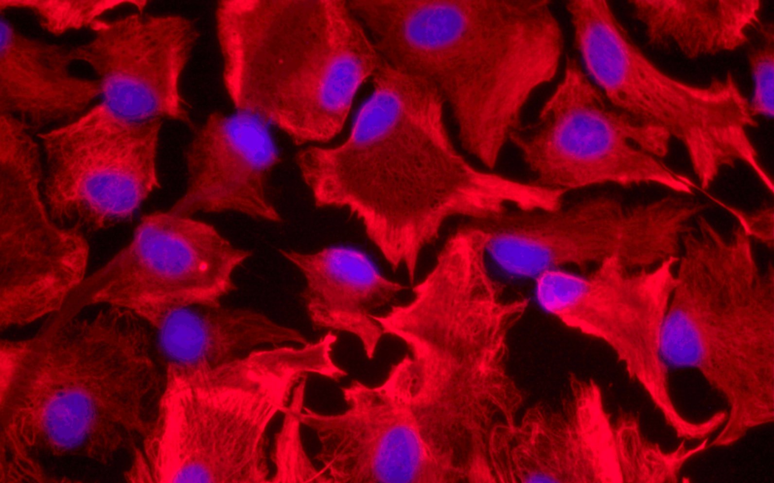
Astrocytes stained with anti-GFAP (Glial fibrillary acidic protein; astrocyte's specific protein).
Data obtained by Mélanie Tremblay.

In order to reproduce hepatic encephalopathy pathology, we use either in vitro and in vivo models.
Astrocytes are very abundant glial cells in the brain. It exist some astrocyte cell lines, but these are usually derived from cancer cells, which may not reflect the non-pathogenic situation. Instead, we use primary cells, which we isolate from rat brains. By culturing them, we can study effect of a pathogenic factor.
Endothelial cells are the cells forming the walls of blood vessels. Because the blood-brain barrier is a crucial structure for the health of the brain, we use primary rat endothelial cells to study the particular roles of this type of cell.
Astrocytes stained with anti-GFAP (Glial fibrillary acidic protein; astrocyte's specific protein).
Data obtained by Mélanie Tremblay.
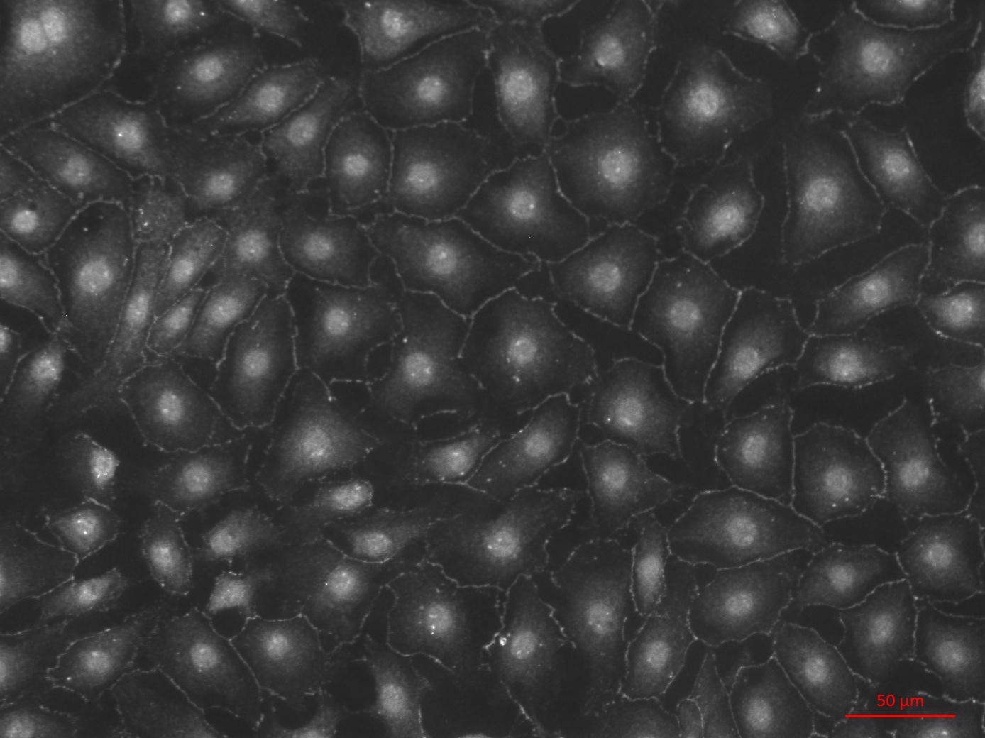
Brain endothelials cells stained with anti-ZO-1 (Zona-occludens-1; BBB specific protein).
Data obtained by Mariana Oliveira.
Over time many different animal models have been developed to study HE. Among those, we works mostly with two surgical rat models showing many characteristics of chronic liver disease.
Bile-duct ligation is a surgery causing the destruction of the liver, equivalent to a biliary cirrhosis. Normally, the liver synthesizes and excretes bile though the bile-duct into the small intestine. However, the bile-duct ligation surgery blocks this excretion. This in turns accumulate the bile in the liver which destroys the liver due to the bile’s toxicity. Thus, the bile-duct ligation model provokes liver fibrosis that eventually becomes cirrhotic. Animals develop hyperammonemia, jaundice, ascites, cerebral edema, portal hypertension, dysfunction of the immune system and minimal HE. These models develop motor and memory difficulties.
Portocaval anastomosis is a surgical procedure by which systematic circulation is diverted from the liver. This model mimics the creation of collateral circulation during cirrhosis. Normally, the blood coming from the intestines enters the liver via the portal vein, is detoxified and goes out through the hepatic veins to feed the body's organs. In order to remove the liver's contribution to blood circulation, the portal vein is ligated and sutured to inferior vena cava.
This model has been recognized as being similar to hepatic encephalopathy, though with some abnormalities in behavior. It ultimately leads to hyperammonemia. However, no cerebral edema or Alzheimer type 2 cells have been detected.
It involves either hepatic devascularisation by diverting and stopping the liver’s circulation or by total hepatectomy, a complete removal of the liver.
It requires the use of hepatotoxic substances such as toxins or drugs to produce acute liver failure. Examples : thioacetamide (TAA), acetaminophen (APAP).