Bobjgalindo (https://commons.wikimedia.org/wiki/File:MRI_brain_tumor.jpg), https://creativecommons.org/licenses/by-sa/4.0/legalcode

Cerebral edema is an abnormal accumulation of fluid in the cerebral tissue, intercellularly and intracellularly in astrocytes.
The causes of cerebral edema are very varied: they can result from a mechanical cause (cerebral trauma), a metabolic disorder (accumulations of toxic derivatives) but also following a cardiovascular accident, secondary to cerebral hemorrhages, tumors, allergy, sepsis or hypoxia.
Liver diseases, acute and chronic, are also a cause of edema.
There are several types of cerebral edema: vasogenic, cytotoxic, osmotic and interstitial.
Depending on the severity of the edema, the underlying symptoms may be more or less severe. The pressure of the cranial box on the cerebral tissues causes a compression of the veins, arteries and nerves. This intracranial hypertension has an effect on the cerebral perfusion resulting in local necrosis and ischemia (decreased oxygen intake).
Symptoms may vary from walking, speech, dizziness, dizziness, vomiting, visual impairment, vigilance or loss of knowledge.
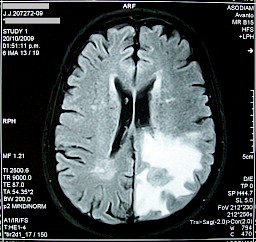
The diagnosis is done by scanning or magnetic resonance imaging (MRI) (allows to visualize the size of an anomaly in the brain tissue).
The treatment of cerebral edema is drug-based corticosteroids with monitoring of intracranial pressure for treatment adaptation. The objective of the treatment is to eliminate excess fluid and decrease the intracranial pressure. Depending on the cause of cerebral edema, it is possible to envisage surgical treatment by setting up a bypass (shunt) in order to draining excess fluid.
Bobjgalindo (https://commons.wikimedia.org/wiki/File:MRI_brain_tumor.jpg), https://creativecommons.org/licenses/by-sa/4.0/legalcode